


 632 / 916

S628
25th European Congress of Psychiatry / European Psychiatry 41S (2017) S583–S644
Disclosure of interest
The authors have not supplied their decla-
ration of competing interest.
http://dx.doi.org/10.1016/j.eurpsy.2017.01.1018EV0689
Cerebellar activity in young people
with familial risk for psychosis — The
Oulu brain and mind study
T. Jukuri
1 ,∗
, V . Kiviniemi
2 , J. Veijola
11
Research Unit of Clinical Neuroscience, University of Oulu,
Psychiatry, Oulu, Finland
2
Oulu University Hospital, Diagnostic Radiology, MIPT, Oulu, Finland
∗
Corresponding author.
Objective
The cerebellum plays a critical role in cognition and
behavior. Altered function of the cerebellum has been related to
schizophrenia and psychosis but it is not known how this applies
to spontaneous resting state activity in young people with familial
risk for psychosis.
Methods
We conducted resting-state functional MRI (R-fMRI) in
72 (29male) young adults with a history of psychosis in one or both
parents (FR) but without their own psychosis, and 72 (29 male)
similarly healthy control subjects without parental psychosis. Both
groups in the Oulu Brain and Mind Study were drawn from the
Northern Finland Birth Cohort 1986. Participants were 20–25 years
old. Parental psychosis was established using the Care Register
for Health Care. R-fMRI data pre-processing was conducted using
independent component analysis with 30 and 70 components. A
dual regression technique was used to detect between- group dif-
ferences in the cerebellum with p b 0.05 threshold corrected for
multiple comparisons.
Results
FR participants demonstrated statistically significantly
increased activity compared to control subjects in the anterior lobe
of the right cerebellum in the analysis with 70 components. The
volume of the increased activity was 73 mm
3
. There was no dif-
ference between the groups in the analysis with 30 components
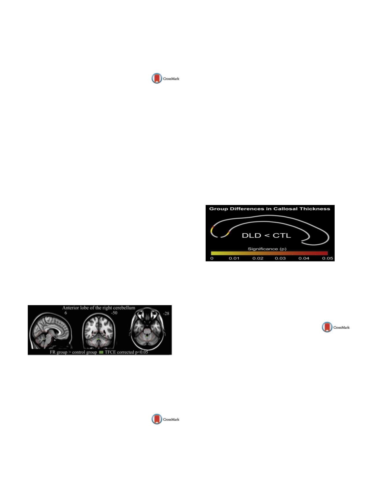
( Fig. 1 ).Conclusion
The finding suggests that increased activity of the
anterior lobe of the right cerebellum may be associated with
increased vulnerability to psychosis. The finding is novel, and needs
replication to be confirmed.
Fig. 1
Disclosure of interest
The authors have not supplied their decla-
ration of competing interest.
http://dx.doi.org/10.1016/j.eurpsy.2017.01.1019EV0690
Atypical callosal morphology in
developmental language disorder
E. Luders
1 ,∗
, F. Kurth
1, L. Pigdon
2, G. Conti-Ramsden
3, S. Reilly
4,
A. Morgan
21
UCLA School of Medicine, Psychiatry, Los Angeles, USA
2
Murdoch Childrens Research Institute, Murdoch Childrens Research
Institute, Melbourne, Australia
3
University of Manchester, University of Manchester, Manchester,
United Kingdom
4
Menzies Health Institute at Griffith University, Gold Coast, Australia
∗
Corresponding author.
Introduction
Developmental language disorder (DLD) is com-
mon, yet the neurobiology of DLD is poorly understood. A key
hypothesis suggests atypical functional lateralization of language,
which might be accompanied structurally by a deficit in inter-
hemispheric connectivity of language-related regions. Indeed,
aberrations of the corpus callosum have been associated with lan-
guage deficits in children with frank neurological lesions and/or
born pre-term. In contrast, studies examining the corpus callosum
in children with DLD remain elusive.
Objective
We aimed to expand this largely understudied field by
comparing callosal morphology between 17 children with DLD and
17 typically developing children carefully matched for sex and age.
Methods
We analyzed high-resolution structural magnetic res-
onance imaging data applying a well-validated computational
approach, which captures the thickness of the corpus callosumwith
a high regional specificity at 100 equidistant points.
Results
As shown in
Fig. 1 ,we observed a significantly thinner
corpus callosum, particularly in the splenium, in children with DLD
compared to typically developing controls (DLD < CTL).
Conclusions
These findings indicating pronounced aberrations in
the brain’s largest whiter matter tract make an important contribu-
tion to an understudied field of research and support the theory that
DLD is accompanied by atypical lateralization of language function.
Fig. 1
Disclosure of interest
The authors have not supplied their decla-
ration of competing interest.
http://dx.doi.org/10.1016/j.eurpsy.2017.01.1020EV0691
Quantitative EEG may help
differentiating bipolar disorder at old
age from frontotemporal dementia
S.Z. Metin
1 ,∗
, B. Metin
2, B. Kocarslan
2, C. Salcini
3, N. Tarhan
11
Uskudar University, Psychiatry, Istanbul, Turkey
2
Uskudar University, Psychology, Istanbul, Turkey
3
Uskudar University, Neurology, Istanbul, Turkey
∗
Corresponding author.
Introduction
Especially the behavioral variant of Frontotemporal
Dementia (FTD) may present with impulsivity, social disinhibition
or depressive symptoms and these symptoms may create a clini-
cal profile very similar to Bipolar Disorder (BD). In clinical practice,
this similarity at symptom level creates substantial diagnostic con-
fusion and often errors. As the treatment approach to the two
disorders differ significantly, it is essential to make a reliable dif-
ferential diagnosis.
Aim
In this study we aimed to identify EEG differences between
FTD and BD.
Methods
For this aim we recruited 22 patients with FTD and 32
patients with BD. Patients in both groups were evaluated with a
standardized neuropsychological battery and structural MRI. All
patients were evaluatedwith resting EEG. Therewere no significant
age and gender differences between groups.