


 353 / 916

25th European Congress of Psychiatry / European Psychiatry 41S (2017) S303–S364
S349
performed the wavelet analysis on EEG recorded during the face
expectation period: 1000–2000ms from the cue onset.
Results
We found the emotional modulation (EM) in EEG
rhythms during the expectation of angry vs. neutral faces in both
groups. Statistical comparison of the spectral power using 2
×
2 fac-
torial design showed that the EMdifferences (
P
< 0.05) between the
groups were in the left parietal locations in 9Hz and in 16–18Hz,
in the right parietal locations in 27–28Hz, and in the right frontal
area in 30–31Hz.
Conclusions
The unconscious expectation of angry vs. neutral
faces resulted in EM differences between the MDD and healthy
controls in the right frontal and bilateral parietal areas mostly in
beta and gamma ranges.
Disclosure of interest
The authors have not supplied their decla-
ration of competing interest.
http://dx.doi.org/10.1016/j.eurpsy.2017.02.321EW0708
Brain pathway differences between
Parkinson’s disease patients with and
without depressive symptoms
S. Mohammadi Jooyandeh
1 ,∗
, T.C. Baghai
1, M.H. Aarabi
2,
M. Dolatshahi
3, B. Langguth
11
University hospital Regensburg, psychiatry and psychotherapy,
Regensburg, Germany
2
Basir eye health research center, neuroscience, Tehran, Iran
3
Tehran university of medical sciences, student’s scientific research
center, Tehran, Iran
∗
Corresponding author.
Introduction
Depression occurs frequently in patients suffe-
ring from Parkinson’s disease (PD). However, the neural basis of
depression in PD remains unclear. Diffusion magnetic resonance
imaging (DMRI) connectometry is based on the spin distribu-
tion function (SDF), which quantifies the density of diffusing
water.
Aim
The aim of this study was to assess the microstructural
changes in the brain connectivity of PD patients with and without
depressive symptoms.
Methods
DMRI was used to assess microstructural abnormali-
ties in the brains of 16 PD patients with depressive symptoms
compared to 11 PD patients without depressive symptoms.
Data used in the preparation of this paper were obtained from
the Parkinson’s progression markers initiative (PPMI) database
( http://www.ppmi-info.org/data/). This dataset was acquired on a
3-Tesla scanner (Siemens), producing 64 DWI at b = 1000 s/mm
2
and one b0 image. Diffusion MRI data were corrected for
subject motion, eddy current distortions, and susceptibility arte-
facts due to magnetic field inhomogeneity. DMRI connectometry
was conducted in a total of 27 patients using percentage
measurement.
Results
PD Patients with depressive symptoms showed decrea-
sed anisotropy (FDR < 0.05) in the fornix bilaterally, left inferior
longitudinal fasciculus (ILF) and corticospinal tract bilaterally com-
pared to PD patients without depressive symptoms.
Conclusions
Lesser WM integrity of the left ILF fibers, which
connect visual face recognition areas to the amygdala and hippo-
campus, seems to be associated with depressive symptoms in PD
patients. Our study supports the hypothesis that neurodegenera-
tive processes in projections from the somatosensory, cingulate,
and insular cortices may be related to depression in PD.
Disclosure of interest
The authors have not supplied their decla-
ration of competing interest.
http://dx.doi.org/10.1016/j.eurpsy.2017.02.322EW0709
Meta-analysis of aberrant brain
activity in psychopathy
T. Poeppl
1 ,∗
, M. Donges
1, R. Rupprecht
1, P. Fox
2, A. Laird
3,
D. Bzdok
4, B. Langguth
1, S. Eickhoff
51
University of Regensburg, department of psychiatry and
psychotherapy, Regensburg, Germany
2
University of Texas health science center, research imaging
institute, San Antonio, USA
3
Florida international university, department of physics, Miami, USA
4
RWTH Aachen university, department of psychiatry and
psychotherapy, Aachen, Germany
5
Heinrich Heine university, department of clinical neuroscience and
medical psychology, Duesseldorf, Germany
∗
Corresponding author.
Introduction
Psychopathy is characterized by superficial charm,
untruthfulness, lack of remorse, antisocial behavior, egocentricity
as well as poverty in major affective reactions. This clinical profile
has been empirically conceptualized and validated. Recent brain
imaging studies suggest abnormal brain activity underlying psy-
chopathic behavior. However, no reliable pattern of altered neural
activity has been disclosed so far.
Objective
To identify consistent changes of brain activity in psy-
chopaths and to investigate whether these could explain known
psychopathology.
Methods
First, we used activation likelihood estimation to
meta-analyze brain activation changes in psychopaths across 28
functional magnetic resonance imaging studies reporting 753 foci
from 155 analyses (
P
< 0.05, corrected). Second, we functionally
characterized the ensuing regions employing meta-data of a large-
scale neuroimaging database (
P
< 0.05, corrected).
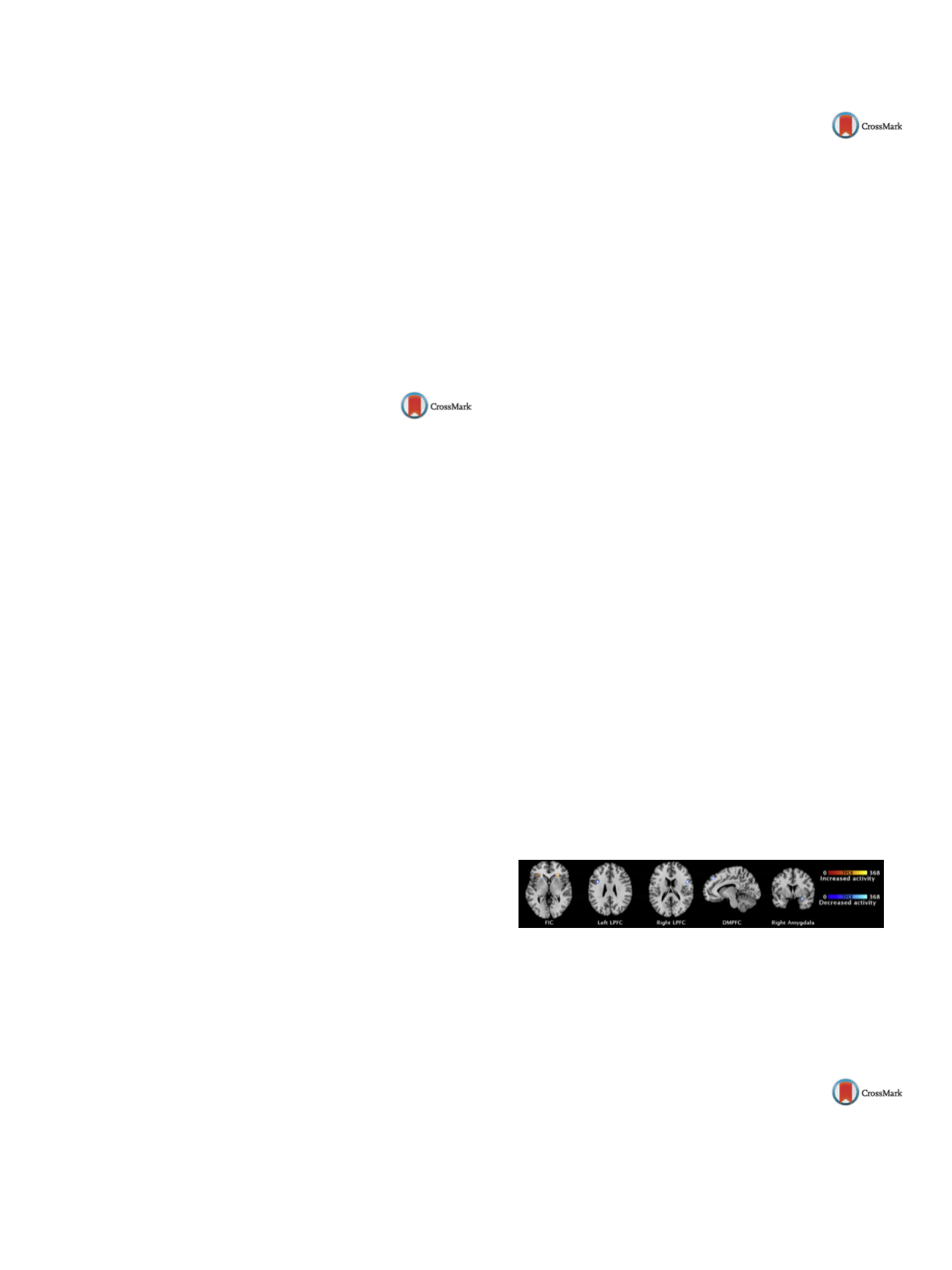
Results
Psychopathy was consistently associated with decreased
brain activity in the right amygdala, the dorsomedial prefrontal
cortex (DMPFC), and bilaterally in the lateral prefrontal cortex
(LPFC). Consistently increased activity was observed bilaterally in
the fronto-insular cortex (FIC)
( Fig. 1 ).Moreover, we found that
the physiological functional role of the candidate regions related
to social cognition (DMFPC), cognitive speech and semantic pro-
cessing (left FIC/LPFC), emotional and cognitive reward processing
(right amygdala/FIC) aswell as somesthesis and executive functions
(RLPFC).
Conclusion
Psychopathy is characterized by abnormal brain acti-
vity of bilateral prefrontal cortices and the right amygdala, which
mediate psychological functions known to be impaired in psycho-
paths. Hence, aberrant neural activity can account for pertinent
psychopathology in psychopathy.
Fig. 1
Disclosure of interest
The authors have not supplied their decla-
ration of competing interest.
http://dx.doi.org/10.1016/j.eurpsy.2017.02.323EW0710
Cannabis use decreases prefrontal
glutamate levels in early psychosis
S. Rigucci
1 , 2 ,∗
, L. Xin
3, P. Klauser
1, P.S. Baumann
1, L. Alameda
1,
M. Cleusix
4, J. Raoul
4, C. Ferrari
1, M. Pompili
2, R. Gruetter
5,
K. Do Cuenod
4, P. Conus
11
Centre hospitalier universitaire vaudois, CHUV, department of
general psychiatry, Lausanne, Switzerland