


 348 / 916

S344
25th European Congress of Psychiatry / European Psychiatry 41S (2017) S303–S364
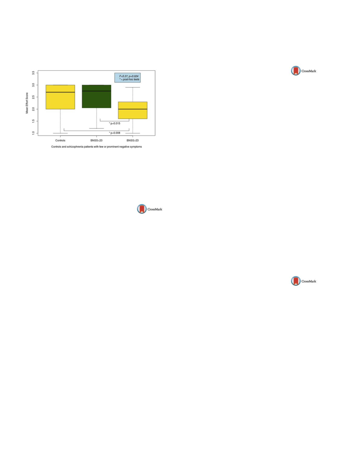
with less negative symptoms and controls
( Fig. 1 ). Our reward
task correlates well with negative symptoms. Thus, it could offer
a behavioral measure of negative symptoms. It could be a good ins-
trument to study the neurobiological basis of negative symptoms
using functional techniques.
Fig. 1
Reward task output in controls and schizophrenia patients
Disclosure of interest
The authors have not supplied their decla-
ration of competing interest.
http://dx.doi.org/10.1016/j.eurpsy.2017.02.308EW0695
Brain connectivity in patients with
schizophrenia related to psychological
stress
M. Castro
∗
, L. Drucaroff , E. Costanzo , A. Wainsztein , S. Guinjoan ,
M. Villarreal
Conicet, Fleni, UBA, psychiatry, Caba, Argentina
∗
Corresponding author.
Introduction
It is commonly accepted that in most patients with
schizophrenia external factors act on genetic predisposition to pro-
duce active psychotic symptoms. In fact, we showed that patients
with schizophrenia have an abnormal brain activation and per-
ipheral autonomic response to psychological stress. We sought to
characterize the brain connectivity networks of such response in
schizophrenia.
Methods
We studied the pattern of brain connectivity in rela-
tion to mental arithmetic stress paradigm in 21 patients and 21
healthy subjects aged 18 to 50 years, using 3T-fMRI. A period of
6minutes of resting state acquisition (PRE) were followed by a
block design with three 1-minute CONTROL task (one digit sum), 1-
minute STRESS task (two digit subtraction) and 1-minute rest after
task (POST). Pairwise Pearson correlationswere calculated between
90 regions of interest. Data were analyzed with MATLAB and SPSS
software.
Results
Patients with schizophrenia showed a lower connec-
tivity network between fronto-temporal limbic areas compared
with control subjects during control and stress task. Moreover,
we observed a great variability of link density during resting state
in patients but not in controls, and it diminishes in response to
task.
Conclusions
Patients present abnormalities in networks rela-
ted to stress response showing an alteration in fronto-temporal
connectivity, and a poor and random modulation of these net-
works at rest. Current and previous findings suggest abnormal
fronto-temporal connectivity that ultimatelywould lead to psycho-
tic symptoms emergency in response to an environmental stressor
and, even, could be related to hypervigilance and misattribution
feeding into the paranoid cognition characteristic of patients with
schizophrenia.
Disclosure of interest
The authors have not supplied their decla-
ration of competing interest.
http://dx.doi.org/10.1016/j.eurpsy.2017.02.309EW0696
Non-verbal learning disorder:
Neuropsychological profile and neural
correlates. A structural magnetic
resonance imaging study
M. Cervino
∗
, P. Castrillo , R. Guijarro
Complejo hospitalario universitario de Granada, servicio Andaluz de
Salud, unidad de rehabilitación de Salud Mental, Granada, Spain
∗
Corresponding author.
Non-verbal learning disorder (NVLD) is a neurological condition
which is considered to be a learning disability. It is characterised
by a specific dysfunction in motor, visuospatial and social skills in
patientswith a normal intellect and development of language.War-
ning signs in school are poor psychomotor coordination, arithmetic
skills and drawing activities. Social judgment and social problem
solving are also typically impaired. Furthermore, these patients
seem to have increasing risk of emotional disorders. Most of ima-
ging studies and current theories suggest that a dysfunction of
white matter in the right hemisphere could be the cause. Howe-
ver, there is a lack of consensus among experts regarding whether
NVLD exists and what could be the underlying causes for NVLD
symptoms. The aim of this paper is to clarify the neural corre-
lates underlaying the cognitive functioning of these patients. With
this objective, we analyzed a sample of brains of children with
and without NVLD. We used the structural MRI technique and
the voxel-based morphometry analysis. The diagnosis of the chil-
dren were based on neuropsychological data. The present study
suggests that not only white matter of the right hemisphere is dys-
functional in these patients. Some other gray matter areas such
as precuneus (superior parietal lobule) may also be affected in
NVLD.
Disclosure of interest
The authors have not supplied their decla-
ration of competing interest.
http://dx.doi.org/10.1016/j.eurpsy.2017.02.310EW0697
Apathy in depression: An arterial spin
labeling study
C. Conan
1 ,∗
, J.M. Batail
1, I. Corouge
2, J. Palaric
1, G. Robert
1,
D. Drapier
11
Centre hospitalier Guillaume-Regnier, PHUPA, Rennes, France
2
INRIA, VisAGeS project-team, Rennes, France
∗
Corresponding author.
Introduction
Apathy is usually defined as a lack of goal-directed
behavior. Although it is observed in about 30% of depressed
patients, neurovascular mechanisms underpinning apathy remain
little-known.
Objectives
The main objective of this study was to compare
the cerebral perfusion of apathetic depressed patients with non-
apathetic depressed patients by arterial spin labeling (ASL), a
quantitative and non-invasive perfusion magnetic resonance ima-
ging (MRI) technique. The secondary objectives were to study
their clinical profile and their correlation with cerebral perfusion
data.
Methods
This study was conducted from a cohort of depressed
patients in Rennes, France. Eighty-three depressed patients were
included, of whom 22 were apathetic (AES
≥
42), 61 non-apathetic
(AES < 42). Everyone got a clinical evaluation with scale scree-
nings, especially for apathy (AES), anxiety (STAI) and anhedonia
(SHAPS) as well as a cerebral MRI, including a pseudo-continuous
ASL sequence.